PET

The ADNI PET Core has the responsibility of standardizing the acquisition, quality control, preprocessing and analysis of all PET data collected as part of the ADNI project.

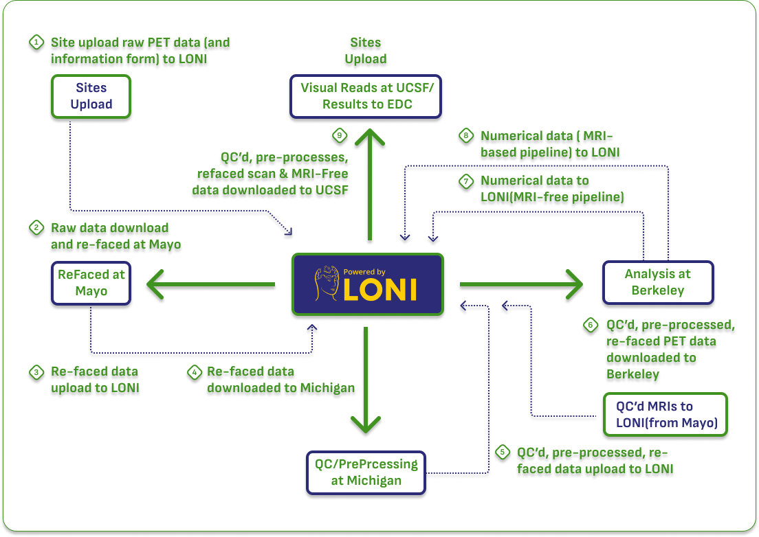
The PET data available to to ADNI data users through the LONI IDA includes:

PET images in DICOM format
PET images in DICOM format - including raw, pre-processed, and post-processed scans.
Numerical summary data in tabular csv format
Numerical summary data in tabular csv format - including regional SUVR measures for both tau and amyloid PET.
Tables containing  detailed acquisition and quality control information
Tables containing detailed acquisition and quality control information for individual scans.
This page provides a high-level overview of the PET component of the ADNI data set. More detailed information is available on the documentation page, and in the PET section of the ask the experts archive.
Investigators who have specific questions for the PET core that are not addressed in any of the resources listed above can submit a question using the ask the experts feature.

Historical PET Availability and Data Types

Understanding what kind of PET data is available on a more granular level can be challenging. ADNI has been active for 20 years, and the types of PET data collected has evolved over time.

This graphic shows which scans were collected at different times across the different phases of ADNI, and which PET tracers were used during different periods.

scans collected at different times across the different phases of ADNI

During each phase of ADNI, different participant groups were studied at different intervals. The following table provides this information:

participant groups were studied at different intervals

Current ADNI4 Methods

In ADNI 4, multiple amyloid and tau PET tracers are used for imaging. PET tracers, with times of acquisition and injected doses are:

PET tracers, with times of acquisition and injected doses

All ADNI scanners must be qualified through a process available in the PET Technical manual. Once a scanner is qualified it does not need re-qualification unless there are substantial changes to hardware or software.

Image Pre-Processing

Images are uploaded to LONI and undergo pre-processing at the University of Michigan to produce the following output of different image types:

1. Co-registered Dynamic

Each 5-minute frame in native space (de-faced, when applicable, or original, raw scan) is co-registered to the first extracted frame of the raw image file in order to motion correct the images.

2. Co-registered, Averaged

Individual 5-minute frames in the dynamic image set are averaged to produce a single image.

3. Co-reg, Avg, Standardized Image and Voxel Size

Each tracer type's first scan (amyloid and tau separately) is reoriented into a standard 160×160×96 voxel image grid with 1.5 mm3 voxels using rigid body registration to achieve standard AC-PC alignment. Subsequent scans are co-registered to their first scan's AC-PC alignment for consistent spatial orientation. Cerebellar gray matter is used as the reference region for SUVr normalization. Importantly, this initial intensity normalization is later replaced (“divided out”) by tracer-specific reference regions in BPIP.

4. Co-reg, Avg, Std Img and Vox Siz, Uniform Resolution

The standardized images are smoothed to a common resolution using scanner-specific 3D-Gaussian filters derived by the University of Michigan team using Hoffman phantom scans carried out at each site11. The effective resolution was selected based on the lowest resolution scanners in ADNI, with resolutions initially set to 8mm3 FWHM and, in 2023, adjusted to 6mm3 FWHM to reflect the lowest resolution of the current scanners. In 2023 all Aβ and tau ADNI PET data were re-processed by UC Berkeley retrospectively at 6mm3 in order to generate a harmonized dataset at the new resolution. Resulting preprocessed PET DICOMs have a standardized voxel size and spatial resolution.

After undergoing pre-processing, images are analyzed at UC Berkeley using in-house software which uses native space, contemporaneous MRI scans that are segemented and parcellated using FreeSurfer. These MR images are coregistered to the PET data, and used to provide PET measures in the Desikan-Killiany atlas. These data are available as csv files through LONI and include extensive methods documentation.

De-facing, quality control, and data flow

ADNI Images undergo extensive processing that includes de/re-facing, done in order to preserve privacy and assure that images cannot be used to identify individuals.

Images undergo multiple steps for quality control including a “pre-check” at Michigan, a full QC evaluation at Michigan, and QC of all data output at Berkeley. Amyloid images are made available, along with the quantitative metrics, to UCSF where they undergo visual reads and these results are provided to the site to release to interested participants. The flow of a single (amyloid) PET image is documented here:

Pre-processing workflow from LONI
2024 Alzheimer’s Disease Neuroimaging Initiative
This website is funded by the Alzheimer’s Disease Neuroimaging Initiative